-
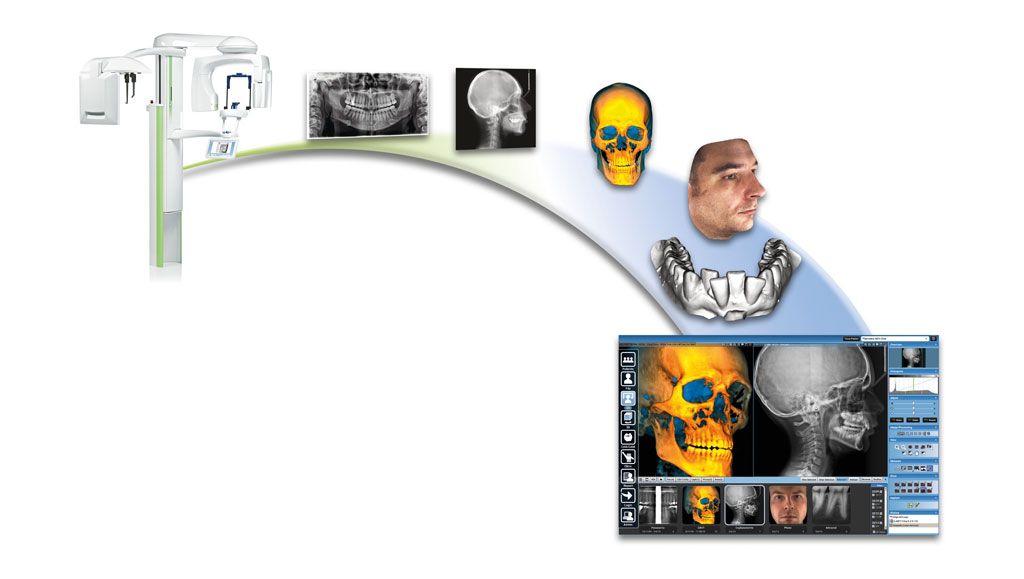
2D / 3D X-ray diagnostics and facial scan in maxillofacial surgery
X-Ray diagnostics
X-Ray diagnostics
2D / 3D X-ray diagnostics in dentistry
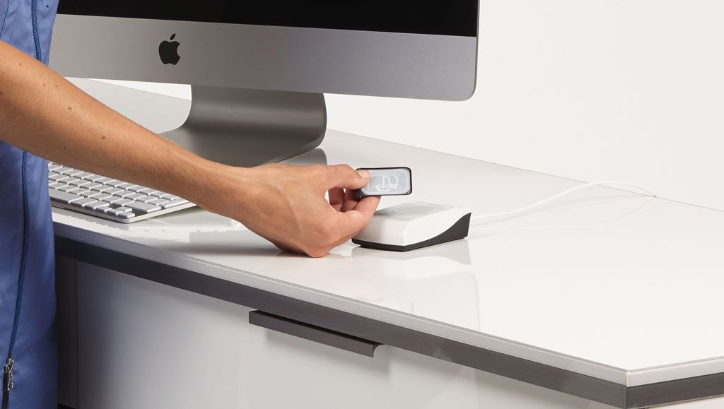
X-ray diagnostics in dentistry
X-ray diagnostics have always been standard in oral and maxillofacial surgery and dentistry. Conventional X-rays show a two-dimensional image of the conditions in the oral cavity. These 2D X-ray images still represent an important basis for the assessment of teeth and jaws.
However, X-ray diagnostics in dentistry has developed enormously in recent years and digital volume tomography (DVT), a very modern and efficient procedure, has been available for some time with which 3D images of teeth and jaws are also possible. 3D X-ray opens up far-reaching possibilities for planning complex dental and maxillofacial surgery.
In our specialist practice for oral and maxillofacial surgery in Berlin Mitte we have both conventional 2D X-ray technology and a digital volume tomograph for the acquisition of 3D X-ray images as well as many years of extensive experience in X-ray diagnostics. Which imaging procedure is used depends on the individual situation of each patient. Depending on the initial situation, we decide when a 2D image and when a 3D X-ray image is required.
When do 2D X-rays make sense?
In many cases, the doctor does not need 3D images to make a reliable diagnosis and plan the treatment. Conventional 2D X-rays are therefore sufficient in most cases. We also rely on digital methods for 2D X-rays, in which the radiation exposure is about 80% lower than with classical, analogue X-ray methods.
Advantages of the digital 2D X-ray procedure
- Within seconds we receive a digital image of your teeth.
- This allows an immediate discussion of the findings with you.
- Individual image sections can be displayed enlarged.
- Both single shots and panorama shots are possible.
- Contrast and brightness of the images can be modified.
- We find caries in the early stages.
- We can judge tooth pockets.
- The position of wisdom teeth can be determined and root canals can be shown.
- Leaks at the crown margin or filling margin can also be clearly identified.
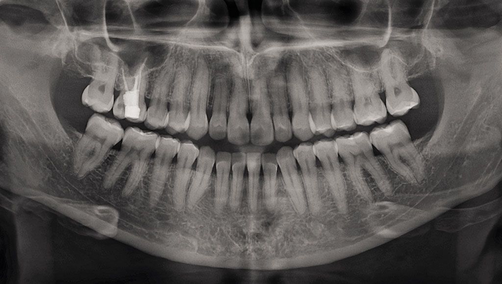
In order to increase the expressiveness of digital 2D images, several images are created in places from horizontal or vertical angles. This results in additional information and a better assessment of the situation.
Intraoperative result and quality control
Digital X-ray during the procedure
Digital imaging procedures are not only used for the preparation of findings, they can also be used during the actual procedure and provide the surgeon with important real-time information. The intraoperative result and quality control during the operation is meaningful and effective, because corrections can be made during the operation instead of after the operation. With the help of digital X-rays, the specialist can, for example, check whether an inserted implant is in the position planned in advance.
Radiation exposure from X-rays
In normal life we are exposed to natural X-rays every day. It is estimated that the average natural radiation exposure of a human being is about 300 μSv per year. With conventional X-rays, the radiation exposure is between 5 and 20 μSv, depending on the exposure. In comparison, the radiation exposure during a flight from Frankfurt to Gran Canaria is between 10 – 18 μSv.
When are 3D process used?
Conventional, digital X-ray images show a 2D image and are a projection of the structures in the beam path. In many cases, 2D imaging procedures are sufficient for the preparation of findings, but not always. In the case of a 2D X-ray image, the third assessment level is missing and it is up to the experience of the treating specialist alone to identify the location and extent of the problem. This is exactly where 2D X-ray diagnostics reaches its limits and questions that can only be solved by a more precise, spatial representation remain unanswered.
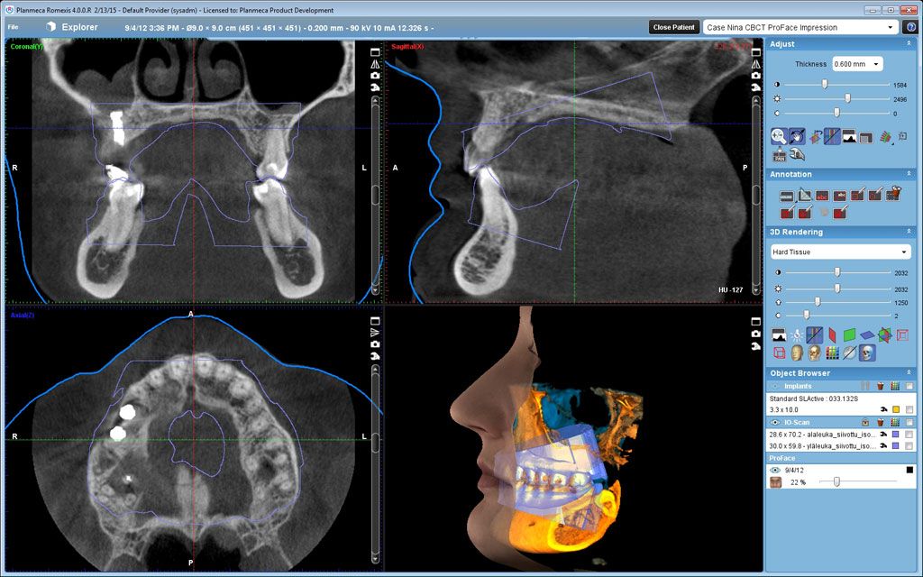
A 3D representation, as made possible by digital volume tomography, offers a more reliable basis for diagnostics. The anatomical conditions are depicted in detail, true to scale and spatially. Whenever the 2D procedure is not sufficient or does not deliver the desired results, DVT is recommended.
Why are 3D images not always created?
If digital volume tomography delivers 3D images of the jaw and teeth and thus provides a very good basis for diagnostics, why is it not always used?
The answer can be found, among other things, by comparing the radiation exposure of both devices. The radiation exposure with a conventional, 2D X-ray machine is still lower than with digital volume tomography. There must therefore be an important reason for using digital volume tomography. The guideline states that a DVT can be prescribed if no clear statement is possible on the basis of the 2D images.
In addition, not every dental practice has a digital volume tomograph. These devices are expensive to purchase and are not yet part of the standard dental equipment. Dentists who do not have a digital volume tomograph can refer their patients to a practice with the appropriate equipment if required.
We have a digital volume tomograph of the latest design as well as the qualification and many years of experience to evaluate the images and develop structured and precise treatment concepts.
What exactly is DVT?
Digital volume tomography (DVT) is an ultra-modern 3D-based X-ray procedure with which spatial images of the skull, teeth and jaw are possible. DVT technology is used in oral and maxillofacial surgery, dentistry and the ENT field.
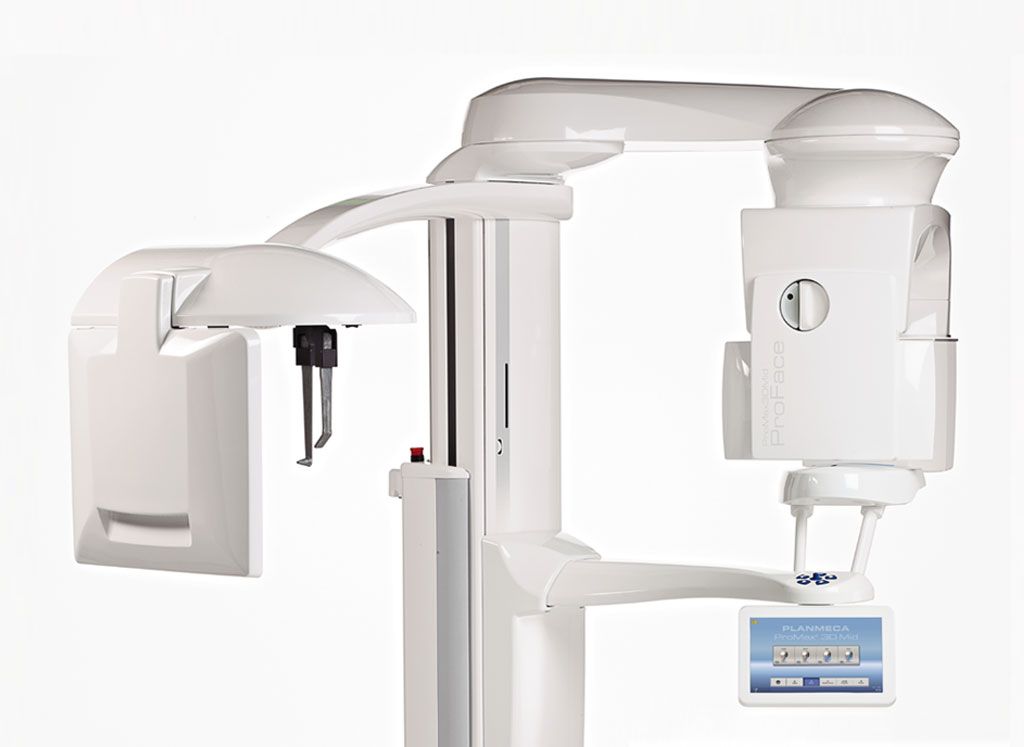
Various structures such as teeth, bones, tooth roots, maxillary sinuses, temporomandibular joints and nerves can be measured and evaluated in detail. In addition to the jaw and temporomandibular joints, we can also visualise part of the paranasal sinuses. Depending on the device, this section can be smaller or larger. For special applications, for example in children, we can reduce the radiation exposure by a fast-scan procedure.
Similar to magnetic resonance tomography and computer tomography, DVT produces sectional images. The DVT process is based on a rotatable mounted X-ray tube. This is opposed by an image sensor. By rotating around the patient’s head, many (approx. 200-400) 2D images are taken. The computer combines these images into a 3D result. The information content of 3D X-ray images is many times higher than that of 2D images.
Diagnosis and therapy become much more precise with this imaging procedure.
Advantages of DVT technology
- Digital volume tomography (DVT) provides clearer and more detailed images compared to conventional 2D X-ray images.
- The design of the DVT device is open, only the patient’s head is x-rayed, which significantly increases the acceptance of this procedure compared to CT (computed tomography) images, where the examination takes place in a tube.
- In contrast to computer tomography, DVT does not require the injection of contrast agents.
- The pure recording time for the DVT is approx. 10-20 seconds. This is a clear advantage over CT recordings, which take about 15-20 minutes.
- Digital volume tomography eliminates the need to visit a radiologist, resulting in greater patient comfort and shorter treatment times.
- Disturbing shadows due to metals (in fillings, bridges, dental crowns made of amalgam and gold) occur to a much lesser extent in comparison to CT images with DVT. This increases the informative value of the 3D images.
- Dental implantations and oral surgical procedures can be simulated and precisely planned with the help of DVT X-rays. This makes treatment results predictable.
- For demanding operations in areas of the maxillary sinus or the mandibular nerves, 3D X-ray technology offers the highest possible level of safety for gentle treatment.
- If one compares the radiation exposure of CT images with the radiation exposure of DVT images, the radiation exposure of DVT images is reduced by approx. 70-90%.
DVT in implantology
An important field of application for digital volume tomography is implantology. As a special practice for implantology, we use DVT already in the diagnostic phase before the actual procedure to clarify the position of various structures, to plan and simulate the procedure. Three-dimensional images can be used, for example, to determine before the procedure whether the bone is high enough and thick enough for the implant to be inserted. The DVT image can thus indicate that bone augmentation is necessary. The exact implant position can also be determined with the DVT images. Good planning and preparation of the actual implantation make the procedure minimally invasive and complications are minimised. At the same time, thanks to this imaging procedure, we can include you as a patient in the planning of your dental prosthesis and thus consider your wishes and ideas even more precisely.
Is every DVT device the same?
When talking about DVT devices, the higher radiation exposure compared to conventional 2D X-ray technology is often referred to as a disadvantage. Manufacturers have responded by developing low-dose modes. As a result, digital volume tomography reaches dose ranges that were previously only known from 2D methods. For this reason, there may well be differences in the DVT devices used, depending on the practice. More modern devices usually also deliver even better and more detailed image quality and thus even more accurate results.
In our practice for oral and maxillofacial surgery, we use state-of-the-art DVT technology of the latest design with integrated low-dose modes. This enables us not only to deliver the best and highest resolution image quality, but also to keep radiation exposure to a minimum.
What are the costs for a digital volume tomography?
As a rule, the costs for DVT are not covered by statutory health insurance. In exceptional cases, the costs can be covered on the basis of a special medical indication. It is therefore a private medical service. The costs for the DVT are regulated by the private medical fee schedule GOÄ. The costs can vary depending on the scope of the examination. Please contact us, we will be happy to advise you on the expected costs of a DVT recording. If you have a private health insurance or a dental additional insurance, you can count in many cases on a cost assumption, which presupposes medical necessity. Should you require confirmation of the medical indication, please do not hesitate to contact us. As an alternative to DVT, a CT (computer tomography) can also be performed by a radiologist.
Are you interested in digital volume tomography or would you like general advice? As a specialist practice for oral and maxillofacial surgery, we are your contact partner.
This might also be of interest to you
Dr. med. Sven Heinrich
Specialist for oral and maxillofacial surgery
plastic and aesthetic operations
– Focus of activities: implantology –
Address:
Friedrichstraße 63
(Entrance Mohrenstraße 17)
10117 Berlin
Opening hours
Mo. to Thu.:
08:00 – 18:00 o’clock
Friday by arrangement
Contact us
Phone: +49 (0) 30 / 84 52 48 88
Email: post@dr-heinrich.berlin
Appointment: arrange here


